

PROJECT OVERVIEW
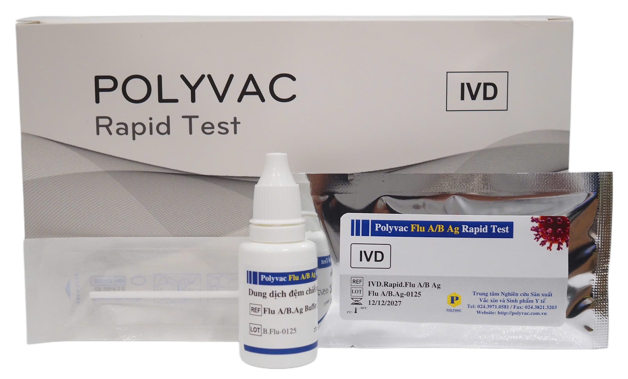
The product "Polyvac Flu A/B Ag Rapid Test" was developed at the Center for Research and Production of Vaccines and Biologicals (POLYVAC).
Research Details
- TOPIC NAME Polyvac Flu A/B Ag Rapid Test
- Classification Product Under Development
General Information
What is the Polyvac Flu A/B Ag Rapid Test?
- Influenza is an acute respiratory infection caused by influenza viruses of the Orthomyxoviridae family. Common symptoms include fever, body aches, chills, chest discomfort, cough, and headache. According to statistics, influenza epidemics cause 3-5 million severe cases and approximately 250,000-500,000 deaths annually worldwide. In Vietnam, over the past 10 years, between 1 million and 1.8 million influenza-like illness cases have been recorded annually, primarily caused by influenza A/H3N2, A/H1N1, and influenza B strains.
- Several diagnostic methods for influenza virus detection have been used, including fluorescence assays, nucleic acid-based tests (NAT), and immunochromatography. This rapid test detects the presence of influenza A/B virus antigens in nasopharyngeal swab samples, supporting the definitive diagnosis of influenza virus infection.
Indications
Polyvac Flu A/B Ag Rapid Test is a qualitative influenza A/B virus antigen test based on immunochromatographic principles to detect influenza A/B antigens present in human nasopharyngeal swab samples. The product is used to support the diagnosis and detection of influenza A/B virus infection.
Operating Principle
- Mouse anti-influenza A virus antibodies and mouse anti-influenza B virus antibodies are simultaneously conjugated with colloidal gold nanoparticles and adsorbed onto the conjugate pad. Mouse anti-influenza A virus antibodies are immobilized at the test line position (T1) of the test device. Mouse anti-influenza B virus antibodies are immobilized at the test line position (T2). Goat anti-mouse IgG antibodies are immobilized at the control line position (C).
- When a sample containing influenza A virus antigen is added to the test device, it forms an influenza A antigen-antibody-gold nanoparticle complex. This complex migrates along the nitrocellulose membrane by capillary action to the test line (T1), where it is captured by mouse anti-influenza A virus antibodies, forming an antibody-antigen-antibody-gold nanoparticle complex that produces a pink-purple line, indicating a positive result. Similarly, for influenza B, when a sample containing influenza B virus antigen is added to the test device, it forms an influenza B antigen-antibody-gold nanoparticle complex. This complex migrates along the nitrocellulose membrane by capillary action to the test line (T2), where it is captured by mouse anti-influenza B virus antibodies, forming an antibody-antigen-antibody-gold nanoparticle complex that produces a pink-purple line, indicating a positive result.
- The remaining conjugate (antibody-gold nanoparticles) not captured by mouse antibodies continues to migrate to the control line and is captured by goat anti-mouse IgG antibodies, producing a pink-purple line that confirms the test is functioning properly.
- Thus, when both pink-purple lines appear at the test line and control line, the sample is positive for influenza virus; when only one pink-purple line appears at the control position, the sample is negative for influenza virus.
Materials Required But Not Provided
- Timer
- Pen
- Tube rack with filter cap
- Gloves
- Face mask
- Pipet
- Pipette tips
Warnings and Precautions
- Read the instructions carefully before performing the test. Incorrect procedures may lead to inaccurate results.
- Check for integrity before use. Do not use the test kit if the foil pouch is punctured, torn, or missing the desiccant sachet.
- Allow all test components to reach room temperature before use.
- Remove the test device from the pouch only when ready to perform the test.
- Do not reuse the test kit.
- Do not use the test kit after the expiration date.
- Do not use the test kit if it has not been stored according to instructions.
Storage
- Store unused test devices and product components at 2-30°C.
- When stored at 2-8°C, ensure that test devices and components are brought to room temperature before use. Do not freeze or expose the device to temperatures above 30°C. Do not use the device beyond the expiration date printed on the packaging.
Sample Collection and Preparation
To prepare the specimen from nasopharyngeal swab, follow this procedure:
- Step 1: Place the extraction tube with filter cap on the tube rack. Add 17 drops (~500 µL) of extraction buffer solution from the buffer bottle (15 mL) into the extraction tube with filter cap.
- Step 2: Insert a sterile swab into the patient's nostril until it reaches the posterior nasopharynx surface. Gently rotate the swab against the posterior nasopharynx surface.
- Step 3: Carefully remove the swab from the nasal cavity. Insert the nasopharyngeal swab into the extraction buffer tube. Rotate the swab at least 5 times. When removing the swab, squeeze both sides of the tube to extract all liquid from the swab.
- Step 4: Securely attach the filter cap to the tube.
Test Procedure
- Step 1: Ensure the specimen and test device are at room temperature before testing.
- Step 2: Open the foil pouch, remove the test device, and place it on a flat surface.
- Step 3: Add 7 drops of sample (120 µL) into the sample well of the test device.
- Step 4: Start the timer.
- Step 5: Read the results within 10 minutes after adding the sample. Do not read results after 10 minutes as accuracy may be compromised.
Quality Control
The control line (C) is integrated into the test device to verify the test procedure. Negative and positive control standards are not provided with the test kit. However, if necessary, quality can be verified using negative and positive control samples in the laboratory to confirm the procedure and product performance.
Result Interpretation
1. NEGATIVE: Only one pink-purple line appears at position C, indicating the sample is negative for influenza A/B virus.
2. POSITIVE: When pink-purple lines appear at both positions C and A/B in the result window (two lines), the sample is positive for influenza A/B virus.
3. INVALID: When no pink-purple line appears at position C in the result window.
Positive results should be interpreted in conjunction with clinical presentation and other available data by the physician.
Test Limitations
- A negative result indicates the absence of detectable influenza A/B virus antigen in the sample. However, a negative result does not exclude the possibility of influenza virus infection, as the antigen level in the sample may be below the detection limit of the test.
- A positive result does not exclude the possibility of co-infection with influenza virus antigens from other pathogenic microorganisms.
- Test results should be interpreted in conjunction with other laboratory test results, epidemiological information, and clinical presentation.