
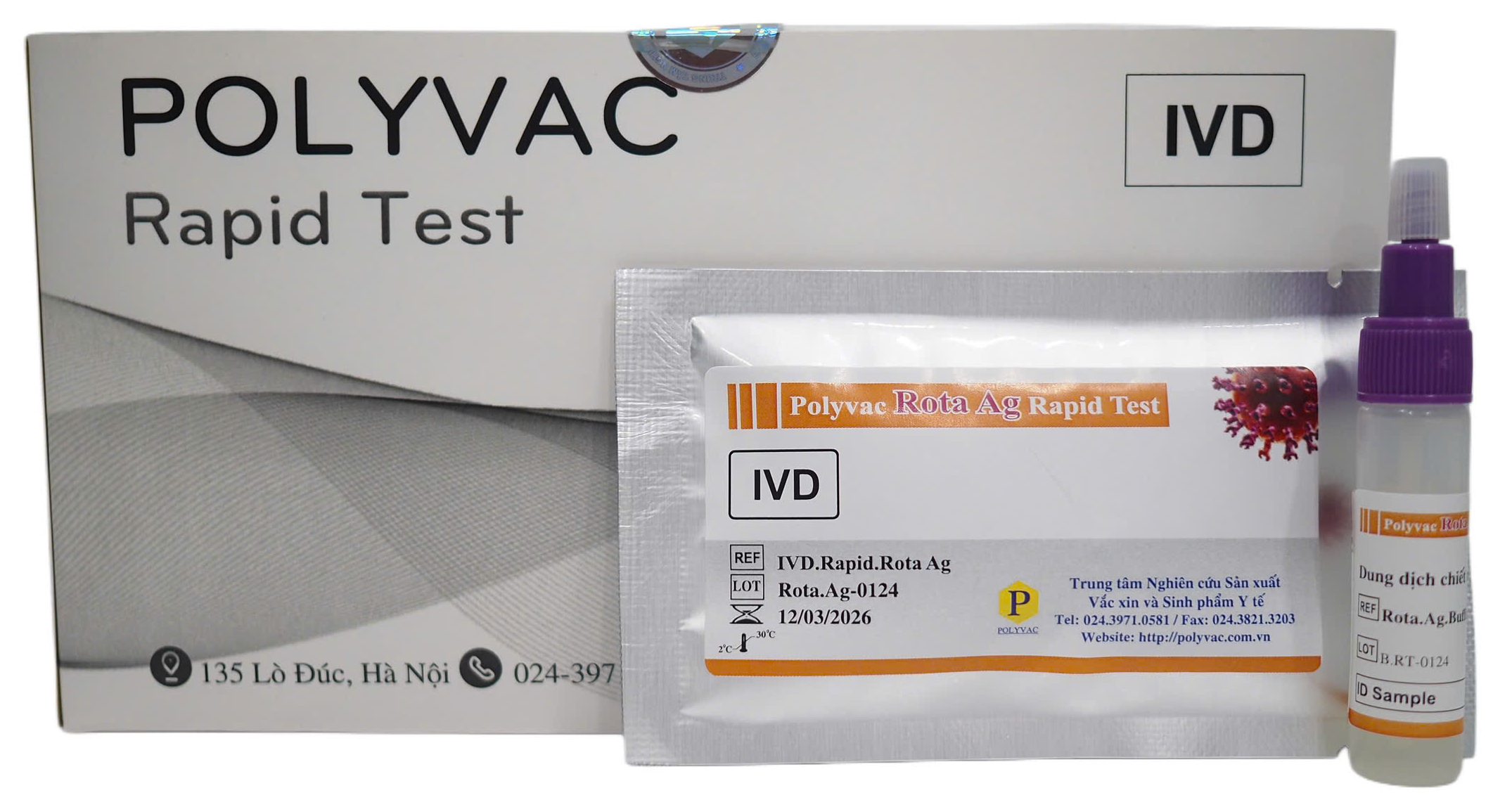
PROJECT OVERVIEW
The product "Polyvac Rota Ag Rapid Test" was developed at the Center for Research and Production of Vaccines and Biologicals (POLYVAC).
Research Details
- TOPIC NAME Polyvac Rota Ag Rapid Test
- Classification Product Under Development
General Information
What is the Polyvac Rota Ag Rapid Test?
- Rotavirus is the leading cause of acute diarrhea in children under 5 years old globally. Each year, this virus causes millions of cases of childhood diarrhea in developing countries, resulting in over 2 million hospitalizations and approximately 500,000 deaths, potentially higher if not diagnosed and treated promptly [1]. Rotavirus comprises multiple genotypes, with G1P[8], G2P[4], G3P[8], G4P[8], and G9P[8] accounting for 90% of Rotavirus diarrhea cases [2].
- Rotavirus infection typically presents with initial symptoms of mild fever, vomiting, and abdominal pain, followed by watery diarrhea lasting 3-8 days [3]. Dehydration occurs more commonly in Rotavirus infections compared to most other infectious agents and is the leading cause of death associated with Rotavirus infection [4]. A patient's stool may contain up to 10¹¹ viral particles. Rotavirus particles remain viable on human hands for at least 4 hours, on dry surfaces for 10 days, and in moist conditions for several weeks.
- Several diagnostic testing methods for Rotavirus infection have been used, including ELISA, latex agglutination, and immunochromatography. This test kit detects the presence of Rotavirus in human stool samples, supporting definitive etiological diagnosis in patients with acute diarrhea.
This section provides detailed information on the operating principle, usage instructions, target populations, and important precautions when using the Rotavirus antigen qualitative test kit. You will find comprehensive guidance on storage, contraindications, specimen collection procedures, and testing protocols.
Operating Principle
- Anti-Rotavirus antibodies are conjugated with colloidal gold particles and adsorbed onto the conjugate pad. Anti-Rotavirus antibodies are immobilized at the test line (T) position on the test cassette. Protein A is immobilized at the control line (C) position.
- When Rotavirus is present in the sample and added to the test device, it forms a Rotavirus-anti-Rotavirus antibody-gold nanoparticle complex. This complex migrates along the nitrocellulose membrane by capillary action to the test line (T), where it is captured by anti-Rotavirus antibodies, forming an anti-Rotavirus antibody-Rotavirus-anti-Rotavirus antibody-gold nanoparticle complex that produces a pink-purple line, indicating a positive result.
- The remaining conjugate (anti-Rotavirus antibody-gold nanoparticles) not captured by the antibodies migrates to the control line and is captured by Protein A, producing a pink-purple line confirming normal test operation.
- Therefore, when both pink-purple lines appear at the test line and control line, the sample is positive for Rotavirus. When only one pink-purple line appears at the control position, the sample is negative for Rotavirus.
Materials Required But Not Provided
- Timer
- Pen
- Sample Container
- Gloves
- Face mask
Warnings and Precautions
- Read the instructions carefully before performing the test. Incorrect procedures may lead to inaccurate results.
- Check for integrity before use. Do not use the test kit if the foil pouch is punctured, torn, or missing the desiccant sachet.
- Allow all test components to reach room temperature before use.
- Remove the test device from the pouch only when ready to perform the test.
- Do not reuse the test kit.
- Do not use the test kit after the expiration date.
- Do not use the test kit if it has not been stored according to instructions.
Product Preparation and Storage Instructions
Store unused test cassettes and unopened stool collection devices at 2-30°C. If stored at 2-8°C, ensure test cassettes and stool collection devices containing extraction buffer are brought to room temperature before use. Do not freeze or expose devices to temperatures above 30°C. Do not use devices past the expiration date printed on the packaging.
Sample Collection and Preparation
To prepare specimens from solid stool samples, follow Procedure A. For liquid stool specimens, follow Procedure B.
Procedure A: Solid Stool Collection
- Step 1: Collect a random stool sample in a clean, dry container.
- Step 2: Open the extraction tube cap, insert the collection swab (attached to the extraction tube cap) into the stool sample at least 5 different locations to collect approximately 10 mg of specimen. Note: For solid stool, confirm that stool has adhered to the grooves on the collection swab; collect from mucoid, viscous areas.
- Step 3: Replace the cap on the tube and tighten securely.
- Step 4: Mix the stool with the buffer by shaking the tube vigorously and wait approximately 20 seconds.
Procedure B: Liquid Stool Collection
- Step 1: Collect a random stool sample in a clean, dry container.
- Step 2: Open the extraction tube cap.
- Step 3: Use the dropper to aspirate the stool sample and dispense 6 drops (100-120 μl) into the extraction tube.
- Step 4: Replace the cap on the tube and tighten securely.
- Step 5: Mix the stool with the buffer by shaking the tube vigorously and wait 20 seconds.
Test Procedure
- Step 1: Ensure specimens and test cassettes are at room temperature before testing. After thawing samples, mix thoroughly before performing the test.
- Step 2: Open the foil pouch, remove the test cassette, and place on a clean, flat surface.
- Step 3: Shake the extraction tube vigorously to ensure the solution inside is homogeneous.
- Step 4: Hold the extraction tube in an upright position. Remove the cap. Add 3 drops of extracted sample (90-120 µL) into the sample well of the test device.
- Step 5: Start the timer.
- Step 6: Read the results after 7-10 minutes. Do not read results after 10 minutes as accuracy may be compromised.
Quality Control
The control line (C) is integrated into the test strip to verify the test procedure. Negative and positive control standards are not provided with the test kit. However, if necessary, quality can be verified using negative and positive control samples in the laboratory to confirm the procedure and product performance.
Result Interpretation
1. NEGATIVE: Only one pink-purple line appears at position C. Result: Negative for Rota virus.
2. POSITIVEWhen pink-purple lines appear at both positions T and C, the result is positive for Rotavirus. The intensity of the T line may vary (darker or lighter) depending on the concentration of Rotavirus in the sample.
3. INVALID: When no pink-purple line appears at position C in the result window.
Positive results should be interpreted in conjunction with clinical presentation and other available data by the physician.
Diagnostic Performance
1. Clinical Performance
412 negative stool samples and 253 positive stool samples were tested using the Polyvac Rota Ag Rapid Test to determine product sensitivity and specificity. Stool samples from patients were also confirmed as negative or positive for Rotavirus by ELISA reference method. Results showed that the Polyvac Rota Ag Rapid Test achieved 95.26% sensitivity and 99.51% specificity.
2. Cross-Reactivity
The Polyvac Rota Ag Rapid Test shows no cross-reactivity with other viruses and bacteria commonly present in stool samples, including Norovirus, Adenovirus, Enterovirus, Escherichia coli, Listeria monocytogenes, Campylobacter coli, Salmonella paratyphi, Salmonella typhimurium, Shigella boydii, Shigella flexneri, Shigella sonnei, Staphylococcus aureus, Vibrio cholerae, Vibrio parahaemolyticus, Candida albicans, Proteus mirabilis (Table 2).
Test Limitations
- A negative result indicates the absence of Rotavirus in the sample. However, a negative result does not exclude the possibility of Rotavirus infection, as this may be due to viral levels below the test's sensitivity threshold or the absence of Rotavirus in the stool at the time of collection.
- A positive result does not exclude the possibility of infection with other gastrointestinal pathogens. Although the test shows no cross-reactivity with the agents listed above, co-infection with other pathogenic microorganisms is possible. Additional microbiological testing should be performed in parallel with this test to rule out other potential causes.
- Test results should be interpreted in conjunction with other laboratory test results, epidemiological information, and clinical presentation.